IAP RAS researchers made a significant contribution to the development of optical coherence tomography (OCT) for imaging the internal structure of biological tissues that are strongly scattering and relatively weakly absorbing media. The research team headed by V. M. Gelikonov and A. M. Sergeev created optical fiber tomographs for noninvasive (endoscopic inclusive) diagnosis of biotissue at a depth up to 2 mm with spatial resolution up to 10 µm. Optical tomograms of the cross-section of surface layers of internal organs in norm and pathology, including cancer, were obtained for the first time. An algorithm was developed for reconstructing parameters of biotissue scattering by OCT images and quantitative description of their state. The information obtained is not provided by other methods of medical diagnosis, including computer tomography and ultrasonography.
|
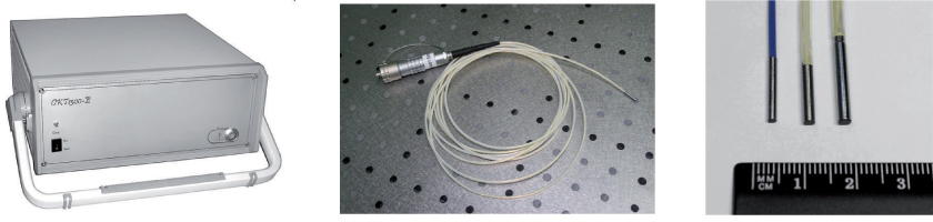
OCT device, endoscopic probe and variants of scanning heads of the developed probes
|
Recent developments enabled increasing the speed and informativity of OCT. Based on known fiber optics and spectral methods an endoscopic modality of OCT device for clinical applications was proposed that operates at a rate of 20 frames per second. A number of endoscopic miniature interchangeable probes were developed that may be used with all standard endoscopic equipment.
A common-path cross-polarization time-domain OCT (CP OCT) has been developed. CP OCT is based on formation of probing light as a pair of orthogonal arbitrarily polarized mutually coherent waves with a definite reciprocal optical delay. The CP OCT image is obtained by detecting in consecutive order of the interference between the waves backscattered both in the initial (co-) and orthogonal (cross-) polarization states with specially designed reference waves. Angular orientation of biotissue axes does not affect the image in the first channel but influences the image in the second channel, allowing ordered layers in the local fragments to be extracted.
|
|
|
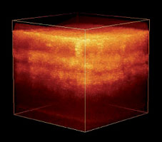
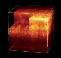
3D-images of finger skin obtained in vivo by spectral OCT at a rate of 20 frames per second
(20 2D-images per second). Left — full view, right — 3D-sections
in arbitrary planes
|
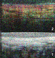
Comparative images of human mucosa in norm in two channels of cross-polarization OCT
|
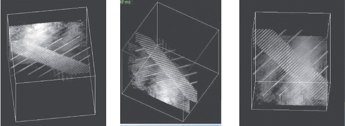
The use of the principles of broadband multiwave digital holography permitted creating a modification of an OCT device without external scanning systems, which opens up new opportunities for updating the method.
IAP RAS has world-class achievements in the area of imaging methods based on low coherence interferometry, including high-sensitivity tomographs for various medical applications.
|
3D images of test structure in the form of a rule with 10 µm step on target glass exposed through a dusty top surface. The surface is above the rule (left), to the left of the rule (center), and under the rule (right)
|