Methods for 3D imaging of biological tissues with cellular and subcellular resolution are becoming a demanded tool in modern neurobiology, developmental biology, and experimental medicine. One of the most popular 3D optical techniques is laser scanning confocal microscopy (LSCM). This method allows imaging biological specimens with subcellular resolution at an up to 200 μm depth and an up to 0.1—0.5 mm field of view. However, a better understanding of structural and functional organization of biological objects, organs and tissues often requires visualization of the cellular structure in a whole organ and even in a whole body. In the recent years such optical technologies of visualizing large optically transparent objects with cellular resolution have been developed.
The most promising of them is fluorescence ultramicroscopy whose principal difference from LSCM is that ultramicroscopy utilizes plane-sheet fluorescence excitation only in a thin layer of biological tissue by means of a special optical system and records fluorescence signals in the orthogonal direction. Such a configuration provides high image contrast due to the absence of unwanted background fluorescence outside illuminated area and prevents direct light from entering the receiving channel of the system. As a result, the point spread function in ultramicroscopy is much less extended in depth. It also gives an advantage of using the recently developed high numerical aperture macroscopic objective lenses with long working distance and large field of view. This allows examining a large area of biological sample with cellular resolution.
A fiber-optic based fluorescence ultramicroscopy setup using a compact single-mode fiber-optic laser radiation transport system with a plane-sheet excitation laser beam up to 20 mm wide and 6—12 μm beam waist was developed at IAP RAS (I. V. Turchin,
V. A. Kamensky, A. N. Morozov) in co-operation with the P. K. Anokhin Institute of Normal Physiology RAMS.
The main advantages of this modification are better focusing parameters due to pure Gaussian shape mode distribution, immunity to external perturbations (temperature, vibrations, and dust), portability, and easy handling. Optical 2D slices and 3D reconstructed images of biological samples (lungs, heart, mouse brain and mouse embryo) were acquired using GFP fluorescence in transgenic mice or autofluorescence.
|
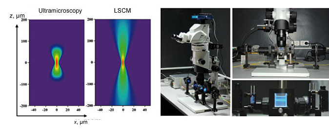
Comparison of point spread functions for fluorescence ultramicroscopy and LSCM (left);
experimental setup for fiber-optic fluorescence ultramicroscopy (right)
|
The system has a high spatial resolution
(5—13 μm) with 6.3x magnification and demonstrates the possibility to investigate large specimens (up to
4 mm) with a single-cell resolution. It can also be used to examine the fluorescent stained samples up to
20 mm in diameter. These parameters surpass the corresponding values in LSCM. The obtained results are important for a detailed study of the structural and functional features of biological tissues in developmental biology, neurobiology and experimental medicine.
|
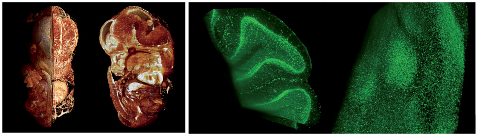
Three-dimensional fluorescence images of optically transparent samples of biotissue obtained
at the fiber-optic ultramicroscopy setup: mouse embryo (left)
and adult mouse cerebellum (right)
|