Fluorescence imaging of experimental animals in vivo has ecently been rwidely used in pre-clinical experimental studies for solution of diverse problems, for example, development of new methods for diagnosis of various pathological processes, assessing efficacy and revealing the mechanisms of drug action, and fast screening of new drugs. In experimental oncology, cancer tumors labeled with fluorescent agents are implanted into animals, which enables in vivo study of the development of primary tumors and metastases at the molecular level. To determine tumor size, the method of epi-illumination fluorescence imaging is generally employed, when the object under study is irradiated by a wide beam of light exciting fluorescence and weak fluorescence is registered from the surface of the animal by a highly sensitive charge-coupled device (CCD). This method allows prompt (1—5 s) assessment of lateral dimensions of a lesion. However, accuracy of this estimate is low for deep-seated tumors, as the light propagated through biotissue is strongly scattered and the image is heavily blurred.
The diffuse fluorescence tomography (DFT) technique provides three-dimensional reconstruction of fluorescent inclusions using information about fluorescence intensity distribution on the surface of the object at different positions of the excitation light source. Unlike the reconstruction in X-ray tomography, the reconstruction of optical inhomogeneities or fluorophore distribution in optical tomography techniques is complicated by multiple light scattering in biotissue. Thus, reconstruction of the fluorophore distribution in DFT demands the development of special algorithms that take into account the features of light propagation in turbid media. A DFT setup with plane transillumination geometry of object scanning was developed at IAP RAS (I. V. Turchin, I. I. Fix,
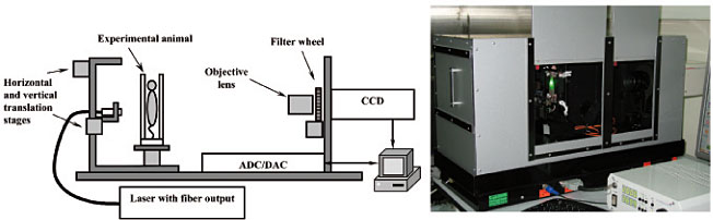
M. S. Kleshnin, V. A. Kamensky). Scanning over the animal's surface is performed by mechanical movement of the optical fiber tip combined with a laser source exciting fluorophore; fluorescence is registered at each position of the source by a highly sensitive CCD with filters blocking the exciting radiation.
|
Scheme and general view of the setup for diffuse fluorescence tomography
|
For reconstruction of a three-dimensional distribution of fluorophore, an iterative algorithm based on Tikhonov functional was developed. The algorithm enables finding a solution to a system of linear equations under the condition of its non-negative components with greater accuracy compared to the solutions obtained by algorithms of a general class. Hybrid models of light transport in tissues developed at IAP RAS are used for solution of a direct problem in DFT. These models permit calculating the illuminated field in a scattering medium at an arbitrary distance from the source with high accuracy using simple analytical solutions of the Radiative Transfer Equation (RTE). For exact solution of the RTE a software package was developed on the basis of the Monte-Carlo simulation winh the graphics processor uswed as a calculator. Results of model experiments on tissue-like phantoms showed a possibility of determining position of the center of fluorescent inclusion and its transverse dimensions to an accuracy not less than 1 mm and in depth size to an accuracy no worse than 1.5 mm. High accuracy of the method was also demonstrated in experiments with small animals in vivo.
The experimental setups created at IAP RAS for fluorescence imaging of experimental animals were tested in the cooperative biological experiments with the Institute of Biochemistry RAS, Institute of Bioorganic Chemistry RAS, Nizhny Novgorod State Medical Academy, University of Nizhny Novgorod, and Russian Oncology Center using various fluorescent agents: fluorescent proteins of different colors, quantum dots, and photosensitizers.
|
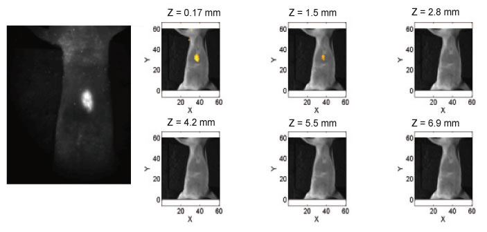
Image of experimental tumor labeled by fluorescent proteins obtained in vivo by surface fluorescence imaging (left)
and reconstructed using DFT (right). Z is the depth of a virtual slice
|