|
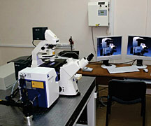
Carl Zeiss laser scanning microscopy complex
LSM 510 META NLO
|
The potential of the fluorescence imaging devices created at IAP RAS was successfully complemented by the Carl Zeiss laser scanning fluorescence microscopy complex LSM 510 META NLO installed at the biophotonics laboratory. The laser scanning microscopy technique for imaging weakly scattering fluorescent objects with submicron resolution is a powerful and constantly upgraded tool for solution of different tasks in fundamental and applied biology. The laser scanning microscopy equipment is used by researchers at IAP RAS in co-operation with the University of Nizhny Novgorod, Nizhny Novgorod State Medical Academy, Institute of Bioorganic Chemistry RAS, and Chemical Physics Institute RAS for in vitro studies of the interaction of fluorescent nanoagents and pharmaceuticals with biological cells (E. A. Sergeeva). These include control of drug delivery to target cells, study of the dynamics of accumulation and subcellular localization of a fluorescent agent at active or passive delivery, and testing new genetically coded sensors based on color fluorescent proteins. It is a preparatory stage for experimental oncology studies executed in vivo.
The LSM 510 META NLO complex includes a femtosecond pulsed laser with tunable wavelength in the near infrared which provides multiphoton excitation of fluorophore and increases imaging depth significantly, even in scattering samples of biotissues (up to 300—500 µm with submicron resolution). In addition, it is possible to observe the structure of unstained collagen-containing biological objects thanks to nonlinear generation of second harmonic of high-intensity probing IR radiation by such structures. The method of second harmonic generation is currently used by researchers at the Nizhny Novgorod State Medical Academy and Moscow State University for investigation of bulk damage of collagen mesh as a result of mechanical, radiation and neoplastic processes.
|
|
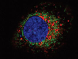
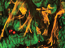
Image of a cell containing fluorescent photosensitizer in lysosomes (top), structure of collagen and elastin fibers
in samples of arterial wall (bottom)
|