Works on development and application of radiophysical methods in medicine were started back in the late nineteen sixties under the supervision of
V. A. Zverev and M. T. Grekhova. They were aimed primarily at the development of acoustic methods of biomedical research, which eventually led to creation of a number of acoustic facilities and techniques. These include ultrasonic echocardiographs for diagnosis of cardiovascular diseases, ultrasonic detectors of gas bubbles in human blood under the action of changing ambient pressure (A. D. Mansfeld, A. M. Reyman), as well as devices for diagnosing the state of biotissues based on vibroacoustic techniques (E. M. Timanin,
V. A. Antonets, V. V. Kazakov).
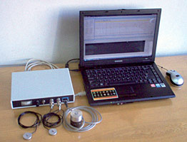 |
Hardware and software complex "Mechanical neuromyograph" comprising a vibration detector for measuring soft tissue elasticity and viscosity, accelerometers for registering motions, an electronic matching unit, and control software
|
Vibration methods of studying human locomotor system: methods of accelerometer diagnostics and vibration viscoelastometry based on recording forced vibratory movements in surface tissues evoked by an external source (E. M. Timanin) have been successfully developed at IAP RAS for a number of years. They are used for development of medical techniques for diagnosis and follow up in surgery, trauma, neurology, sports and space medicine, etc. By combining several techniques a hardware and software complex "Mechanical neuromyograph" for vibration diagnosis of neurological diseases was created.
The complex allows registering vibration signals evoked by involuntary movements of extremities, head, and center of mass of human body (tremorography methods), by test oscillatory movements of extremities (tempography methods), as well as by forced vibratory movements in surface tissues elicited by an external source (viscoelastometry methods). The complex is used to record signals, determine their quantitative parameters and compare them with norm, as well as to form electronic protocols of patient examination.
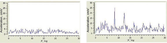 |
Tremor spectra of a healthy person (left) and of a patient with Parkinson's disease
at an early stage (right)
|
Ultrasonic diagnostics based on active ultrasonic location has become a routine examination technique. It allows studying the structure of organs and tissues, blood flow in vessels and heart. However, there is a problem that cannot be solved using this method, namely, measurement of internal temperature of the body. It is known that a change in the temperature of internal parts of the body precedes changes in tissue structure that can be detected by X-rays or ultrasound at later stages of pathology. It is also important to control internal temperature during some therapeutic procedures, especially hyperthermia.
|
|
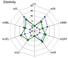
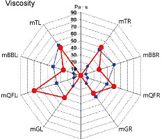
Muscle tone distribution in norm (blue curves) and in a patient with Parkinson's disease (green and red curves).
Muscle group notation: mTL, mTR — m. trapezius; mBBL, mBBR — m. biceps brachii, mQFL, mQFR — m. quadriceps femoris; mGL, mGR — m. gastrocnemius
|
A promising measurement technique is acoustic thermometry that is based on receiving and measuring the intensity of the intrinsic acoustic radiation of the environment generated by thermal motion of its atoms and molecules. The merit of ultrasonic thermo-metry in application to biological tissues is a possibility of using millimeter and submillimeter ultrasonic waves that propagate well in biotissues. This allows directional reception of ultrasonic radiation, thus localizing the heated formation and even reconstructing spatial temperature distribution. Of course, this acoustic emission is very weak, and the range and distribution function of the received noise signal do not differ from those of the receiver noise. However, accumulated signal can be detected and measured by radiometric methods. The attained sensitivity of acoustic thermographs is close to maximum possible and amounts to a few tenths of a degree at measurement time of 5—10 s
(A. D. Mansfeld, R. B. Belyaev, V. A. Vilkov P. V. Subochev, A. G. Sanin, E. V. Krotov).
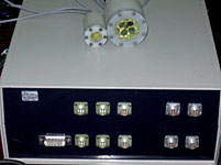 |
Multichannel acoustic thermograph
and its sensors |
In the recent years, the developed devices have been used by physicists in collaboration with medical doctors in a number of experimental clinical studies which demonstrated that it is possible to control internal temperature of the thyroid and mammary glands at laser hyperthermia. These results were obtained jointly by researchers at IAP RAS (A. D. Mansfeld) and IRE RAS (A. A. Anosov) in the Central Clinical Hospital of the Russian Academy of Sciences.
|
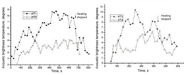
Curves of internal temperature changes during laser hyperthermia of mammary (left) and thyroid (right) glands
|
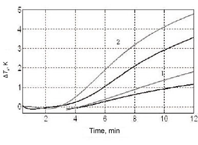
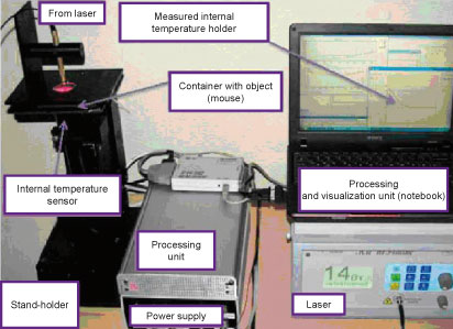
The experiments on laboratory animals carried out jointly with the Nizhny Novgorod State Medical Academy demonstrated the possibility of controlling internal temperature at laser hyperthermia of tumors by injecting into the tumor gold nanoparticles (P. V. Subochev). The efficiency of laser heating of tissue increases
in this case, hence, it is possible to reduce the duration of the procedure. Currently, several types of acous-
tothermographs are under development at IAP RAS that are intended for different tasks of medical diagnostics, including a correlation acoustothermograph capable of extracting a signal from a certain depth, and a multichannel scanning acoustothermograph capable of mapping the field of internal temperatures.
 |
|
Setup for laser hyperthermia in experimental animals
and parallel internal temperature control
|
Increment of acoustic brightness temperature measured during laser hyperthermia by two passive acoustic thermometers: 1 — without nanoparticles, 2 — with injected gold nanoparticles 200—250 nm in size
|